


Before starting an oncology operation, the most important thing a doctor needs is to know as much as possible what they will face.
Is the mass behind the sternum just attached to the pericardium and blood vessels, or has it grown into it? Can it be completely removed? Do I need to get any other treatments first? These questions relate to surgical plans, and some reliable answers often have to wait for post-operative pathological examinations.
The Zhitong Finance App learned that the thymus study in which Deshi (02526) participated is trying to advance valuable information before surgery. This is also the most direct entry point to understanding AI for Science: it allows AI to help researchers process information that is difficult to analyze manually one by one, test the links between images and diseases, and enable more medical problems to be solved and done quickly.
As for this study, the Masaoka-Koga stage in the paper can be understood as a “tumor crossing the boundary map”: stage I is still inside the envelope; stage II begins to break through the boundary, and part of the invasion requires microscopic confirmation; stage III invades neighboring structures such as the pericardium, lungs, or large blood vessels; and stage IV causes dissemination or metastasis.
This map will affect the difficulty of complete resection, preparation for surgery, and treatment sequence. The difficulty is that being “very close” to CT does not mean that the organization has already been violated. The final stage usually relies on surgical and pathologic information.
Patients who can have a complete resection and are in good physical condition usually prioritize surgery; if initial complete resection is difficult to achieve, induction treatment may be performed first and then re-evaluated. Understanding the relationship between tumors and surrounding organs earlier allows doctors and patients to be more prepared.
AI brings another exploration method: it simultaneously calculates the three-dimensional relationship between the texture, shape, and surrounding tissue of the lesion, and learns complex combined characteristics from pre-operative CT and post-operative results. It is difficult for doctors to compare information item by item, so it can be analyzed and tested in batches.
This study was based on iMediimage, combined with imaging omics, to perform phased prediction, 258 training cases, and 65 internal tests in 323 cases of thymic epithelial tumors. The model distinguished two groups, I-II and III-IV. The test accuracy rate was 95.38%, and the AUC was 0.9328. Results were published in the European Journal of Radiology Open in July 2026.
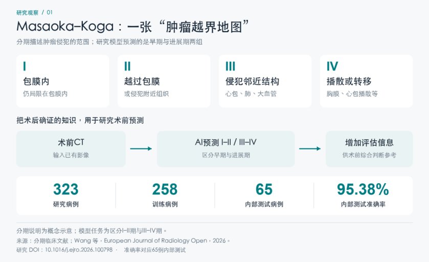
(From post-operative confirmation to pre-operative prediction, the phased task accuracy rate corresponds to 65 internal tests. (Source: Deshi 2026 research papers and clinical literature)
The purpose of this work is to extract new information from existing clinical CT that can help with pre-operative judgment. Once clinically approved in the future, it is expected to add a basis for evaluating resectability, preparing for surgery, and discussing treatment sequences.
The same study also used all 659 cases to distinguish thymic epithelial tumors from other lesions, and WHO pathologic risk groups were performed on 323 cases of thymic epithelial tumors. A set of CT data supports continuous questioning of the nature of the lesion, extent of invasion, and histological risk.
Research like this isn't easy. In 2023, one study traced 10 years of cases from three hospitals and included 373 people for enhanced CT analysis; another study of 187 cases in 2024, segmentation and layer-by-layer correction required about 30 minutes for each case. Behind the data of hundreds of cases is long-term accumulation and extensive professional labor.
With the same CT, AI allows more information to enter calculation, and also allows more medical problems to enter research.
Predicting breast cancer recurrence pushes research into the future. What patients are most concerned about after surgery is whether the cancer will return. Long-term outcomes may be different for patients in the same stage.
Pathological sections show cell and tissue morphology, ultrasound shows the other side of the tumor, and clinical data records the patient's condition. AI puts this information into the same framework, and the system compares a large number of subtle combinations of characteristics and subsequent outcomes, giving researchers an opportunity to find individual differences outside of traditional groupings.
According to Deshi's mid-term results roadshow, the company has conducted 673 studies combining pathology, ultrasound, and clinical information to find links between subtle characteristics and risk of recurrence. The optimized model is 50% off cross-verifying that the C-index is 0.76, which evaluates risk ranking ability. In the future, after receiving clinical approval, such tools are expected to help doctors assess individual risks in more detail and support long-term management and risk communication.
From understanding current disease to predicting future outcomes, AI4S is turning more medical questions into computable and testable research.
The key to morality lies in supporting a common foundation for these studies. iMedImage is first pre-trained on multiple types of images to bring transferable features to new topics; iMedLoop organizes data processing, labeling, training, evaluation, and delivery to reduce the repeated investment of each team in development from scratch.
This can be understood as “first laying a common foundation and then doing specialist training.” Hundreds of cases in the new topic mainly undertake the study of specific tasks; previously accumulated imaging characteristics have become its starting point. Doctors can also focus more on asking questions, judging results, and designing the next round of research.
According to the data disclosed in the company's annual report, a minimum of about 200 images can be used for vertical model development, and the development cycle is 2 to 3 months. iMedMaaS was launched for about 15 months. By the end of June 2026, a total of 158 model projects had been carried out, cooperating with 99 hospitals, covering 61 disease directions.
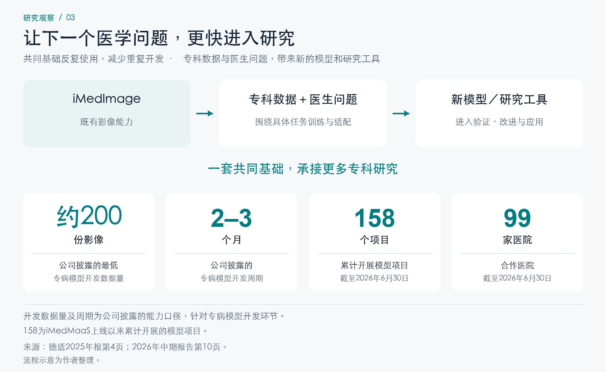
(Common foundation lowers development threshold for new topics)
When the R&D threshold is lowered, limited budgets have the opportunity to support more projects. What is changing is not only the speed of research, but also whether medical ideas can become reality. In the first half of 2026, the service revenue of the Deshi Model was about 94.54 million yuan, an increase of 101.1% over the previous year, and also provided a commercial fulcrum for continuous research and development.
For investors, this means a process that can be continuously expanded: the ability to serve current customers, a common technical foundation supports more topics, and new research provides a source for models and services that can be delivered in the future.
This process is still moving forward. In September 2026, Deshi and the Hong Kong Polytechnic University established a joint laboratory for general artificial intelligence and medical applications, focusing on medical imaging, basic medical models and automation technology, connecting basic research and clinical transformation.
Pushing this path forward, the ideal space for imagination may fall into the prototype of the “medical world model.” The world model learns how the environment changes and how actions affect results; the vision of the medical world model is to deduce the course of the disease based on the patient's condition and further explore the impact of different interventions.
Thymus studies study disease states corresponding to images, and breast research explores future risks indicated by current characteristics. If these capabilities are applied and continue to be connected to follow-up, treatment, and actual outcomes, there is an opportunity to simulate the course of the disease; when the model can reliably learn the relationship between intervention and outcome, doctors can expect to anticipate the changes that the same patient may experience in different treatment sequences.
The most moving imagination of the medical world model is to allow a real diagnosis and treatment decision to be previewed more times in computation.
Diagnosis provides current status, follow-up tests previous predictions, and real results drive model improvement. If such a cycle is formed in the future, there will be an opportunity to explore more problems in computation that are difficult to test in reality.
As the “Godfather of Silicon Valley” Kevin Kelly proposed in a conversation with Desch Chairman and CEO Song Ning, a lasting asset may be the continuous process of generating new medical AI. Deshi's model and platform are providing the foundation for this process.
The scarcity of AI4S is probably hidden here: turning medical problems into new abilities one by one, and then moving towards a larger system of understanding and deducing diseases.